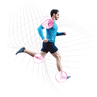
Welcome to the Patient Hub – a space created to help you better understand common injuries affecting the shoulder, knee, foot, and ankle. Here you’ll find educational resources designed to explain the basics of these conditions in simple terms. While this information is not a substitute for medical advice, we hope it supports your conversations with healthcare professionals and helps you feel more informed on your journey.
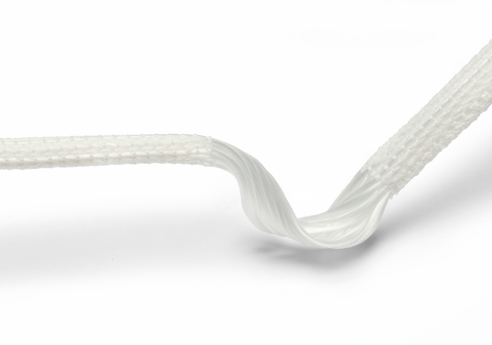
Ligaments are made of fibrous connective tissue and provide stability to the skeleton by connecting one bone to another bone. They are holding joints in place, preventing them from dislocating or injuries and allowing movement such as twisting, jumping, or side-to-side motion. When injured, ligaments are slower to heal than other types of soft tissue due to low vascularity. LARS® is an artificial ligament which mimics the anatomical properties of the natural ligament. Up to now, more than 200 000 LARS® have been implanted globally.
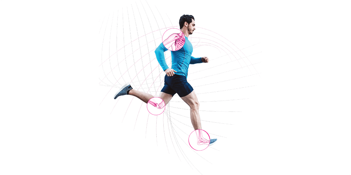
The acromioclavicular joint (ACJ) is a fundamental structure for the stability of the clavicle and of the scapula. Two strong ligaments guarantee its solidity: the conoid and trapezoid ligaments. The acromioclavicular ligament serves to reinforce the joint capsule. Injury of the acromioclavicular joint happens when falling sideways on the shoulder, typical of cyclists, or in strong contrast activities such as rugby.
As the result of a fall or collision, the acromioclavicular joint and surrounding structure can be damaged. The shoulder blade (scapula) is forced downwards, and the clavicle (collarbone) appears prominent. The symptoms are pain, limited motion in the shoulder, swelling, bruising or tenderness at the top of the shoulder.
When experiencing a severe ACJ dislocation/injury, the ligament repair is carried out under general anaesthesia. To stabilize your clavicule, the LARS® is looped around your clavicle and the coracoid process from the shoulder blade. After restoring the relationship between your clavicula and scapula, the LARS® ligament passes through both bones, giving you extreme stability; like a loop it wraps around both bony structures, restoring the anatomy. The new ligament is stabilized by two screws. The procedure will leave you with a little scar of 5 cm running along the top of your shoulder.
The knee joint is stabilized by four ligaments: the anterior and posterior cruciate ligaments (ACL and PCL), the medial collateral ligament (MCL) and the lateral collateral (LCL). Injuries to the ACL and PCL are the most common sports-related injuries, particularly in skiing, basketball, football but also hockey and all contact sports. Without cruciate ligaments, the quality of your knee decreases.
The ACL controls rotation and forward movement of the tibia, while PCL controls its backward movement. When one of these ligaments is injured, people will be unable to jump and land on the knee, accelerate, and then change directions or rapidly pivot on the knee. Being smaller and weaker than the PCL, the ACL is more likely to sustain a complete tear. A damaged ligament results in joint instability, reduced articular performance and high risk to develop osteoarthritis or degenerative changes. The reconstruction of the cruciate ligaments is essential for the return to active life.
Refer to the patient leaflet for additional information and patient rehabilitation.
Ankle ligaments are found throughout your foot, ankle and lower leg. They are short and very strong and is composed of three main ligament complex. The lateral ligament complex is the most injured (lateral ankle sprain). These injuries are typical of sports such as football or athletics, but also of simple falls during everyday life.
Most ankle ligament injuries are caused when the foot twists inwards. The symptoms are pain, swelling and recurrent sprains reduce the capacity of the ligament to properly heal, resulting in long-term ankle instability.
A skin incision is made 4 to 5 cm long. A vertical tunnel is drilled from the tip of the malleolus and emerging from the fibula. By a micro-skin incision, located 2 to 3 cm higher than the previous skin incision, a second tunnel is drilled. The LARS is pulled into the tunnel and fixed using a screw. The suture is made using resorbable threads.
Refer to the patient leaflet for additional information and patient rehabilitation.
This section provides answers to common questions about ligament ruptures and their treatments, helping patients better understand their condition and recovery journey. The information is for general guidance only and should not replace medical advice from a qualified healthcare professional.


When extreme motion is applied to a joint, such as twist, fall or another high-impact event, the ligament can be damaged or totally torn. A damaged ligament results in joint instability, reduced articular performance and high risk to develop osteoarthritis or degenerative changes. Persons with damaged ligaments feel pain, swelling, and discomfort. The activities of daily and professional life could be severely limited by these accidents.
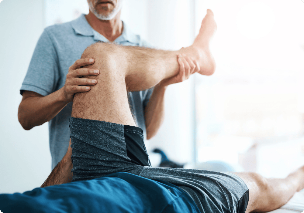
Low grades injuries can heal using a non-surgical treatment, such as a combination of bracing, physical therapy, heat therapy or medication. The healing can take weeks to months. Severe or complete ligament injuries need surgery to restore a natural articulation: the reconstruction or the reinforcement is necessary and autograft, allograft or a synthetic ligament could be used during surgery. The surgical is mandatory to restore a functional articulation, reduce pain, restore circulation, prevent arthritis, and strengthen the surrounding area to support the tissue.
The treatment choice will depend on factors such as your age, your activity level, and the severity of your injury and will be recommended by your orthopaedic surgeon. Tears of any grade require attentive care. Should you suspect a torn ligament, ask for medical advice.
• Autograft: tissue is obtained from another part of the patient’s own body, for example: patellar tendon, hamstring tendon, quadriceps tendon, fascia lata.
• Allograft: obtained from a donated tissue such as a cadaver
• Synthetic/artificial graft: artificially produced and designed to mimic the anatomic ligament fibres
• Hybrid reinforcement: autograft or allograft reinforced with a synthetic/artificial graft
An artificial ligament seeks to mimic the native ligament and is made out of a biocompatible material, functioning in vivo without systemic response in the body. Also, mechanical performances of artificial ligaments can be characterized by abrasion resistance or rotational fatigue limitation. The implantation of an artificial ligament is a very specialized technique, performed in centers of excellence, recommended for athletes and patients requiring high performance.
Note : the medical content of this section have been reviewed by Dr Matteo Rizzo, Switzerland
Please contact us, we look forward to receiving your message!
Movmedix is a leading orthopedic medical device manufacturer in sports medicine with innovative solutions that empower surgeons and enhance patient outcomes.
Movmedix
5 rue de la Fontaine,
21560 Arc-sur-Tille
FRANCE
Tel: +33 (3) 80 37 26 60
Movmedix
5 rue de la Fontaine,
21560 Arc-sur-Tile
FRANCE
Tel: +33 (3) 80 37 26 60